Maharashtra State Board 12th Biology Solutions Chapter 2 Reproduction in Lower and Higher Animals
1. Multiple choice questions
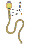
2. Very short answer questions
3. Fill in the blanks
4. Short Answer Questions
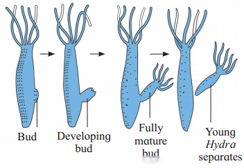
B. Diaphragm, cervical caps and vaults:
2. The site of infection is the mucous membrane in genital, rectal and oral region.
3. Symptoms of syphilis:
5. Answer the Following Questions
1. Menstrual Phase:
2. Proliferative phase/Follicular phase/Post menstrual phase:
(3) Ovulatory phase:
(4) Luteal phase/Secretary phase :
(i) Since the empty Graafian follicle converts itself into corpus luteum under the influence of LH, this phase is called luteal phase in ovary. At the same time, the uterine endometrium thickens and becomes more secretory and hence it is called secretory phase in uterus.
(ii) Corpus luteum secretes progesterone, some amount of estrogens and inhibin. These hormones stimulate the growth of endometrial glands which later start uterine secretions.
(iii) Endometrium becomes more vascularized becomes 8-10 mm. in thickness. These changes are the preparation for the implantation of the ovum if fertilization occurs.
(iv) In absence of fertilization, corpus luteum can survive for only two weeks and then degenerate into a non-secretory white scar called corpus albicans.
(v) If ovum is fertilized, woman becomes pregnant and hormone hCG (human Chorionic Gonadotropin) is secreted by chorionic membrane of embryo which keeps corpus luteum active till the formation of placenta.
2. Expulsion stage:
3. After birth or placental stage : In the last stage of 10 to 45 minutes, once the baby is out then the placenta is also separated from uterine wall and is expelled out as “after birth”. This is accompanied by severe contractions of the uterus.
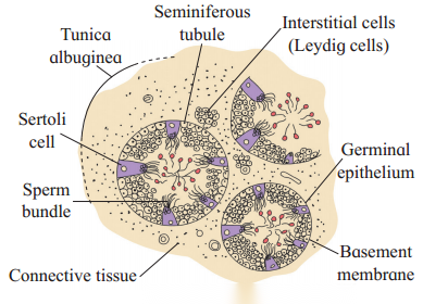
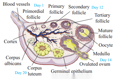
(2) The cortex is covered externally by a layer of germinal epithelium while the medulla contains the stroma or loose connective tissue with blood vessels, lymph vessels and nerve fibres.
(3) Different stages of developing ovarian follicles are seen in the cortex. Each primordial follicle has at its centre a large primary oocyte (2n) surrounded by a single layer of flat follicular cells, then gradually it matures.
(4) In the ovary during each menstrual cycle there is a maturation of primordial follicles into multilayered primary, secondary and Graafian follicles.
(5) Every Graafian follicle has three layers, viz. theca externa, theca interna and membrana granulosa which are from outer to inner side. A space called antrum filled with liquor folliculi is present inside the follicle. There is a small hillock of cells called cumulus oophours or discus proligerus over which the ovum is lodged. The ovum in turn is covered by vitelline membrane, zona pellucida and corona radiata from inner side to outer surface.
(6) Ovarian cortex also shows corpus luteum, or yellow body formed from empty Graafian follicle after ovulation. Corpus luteum is converted into corpus albicans or white body in case of absence of conception.
(2) Coitus Interruptus or withdrawal : In this method, the male partner withdraws his penis from the vagina before ejaculation, so as to avoid insemination. This method also has some drawbacks, as the pre-ejaculation fluid may contain sperms and this can cause fertilization.
(3) Lactational amenorrhoea (absence of menstruation) : This method is based on the fact that ovulation does not occur during the period of intense lactation following parturition so chances of conception are almost negligible. However, this method also has high chances of failure.
(4) Chemical means (spermicides) : In this method chemicals like foam, tablets, jellies and creams are introduced into the vagina before sexual intercourse, they adhere to the mucous membrane, immobilize and kill the sperms.
(5) Mechanical means/Barrier methods:
(i) Condom : It is a thin rubber sheath that is used to cover the penis of the male. Condom should be used before starting coital activity. It also prevents STDs and AIDS.
(ii) Diaphragm, cervical caps and vaults : These devices made of rubber are inserted into the female reproductive tract to cover the cervix diming copulation. They prevent conception by blocking the entry of sperms through the cervix.
(iii) Intra-uterine devices (IUDs) : These are plastic or metal objects placed in the uterus by a doctor. These include Lippes loop, copper releasing IUDs (Cu-T, Cu 7, multiload 375) and hormone releasing IUDs (LNG-20, progestasert). They prevent fertilization of the egg or implantation of the embryo.
(6) Physiological (Oral) Devices : Birth control pills (oral contraceptive pills) check ovulation as they inhibit the secretion of follicle stimulating hormone (FSH) and luteinizing hormone (LH) that are necessary for ovulation. The pill ‘Saheli’ is taken weekly.
(7) Other contraceptives : The birth control implant is similar to that of pills in their mode of action. It is implanted under the skin of the upper arm of the female.
B. Permanent methods surgical operations : In men surgical operation is called vasectomy and in women it is called tubectomy. This method blocks gamete transport and prevent pregnancy.
(ii) Prostate gland:
(iii) Cowper’s glands (Bulbo-urethral glands):
Long answer questions
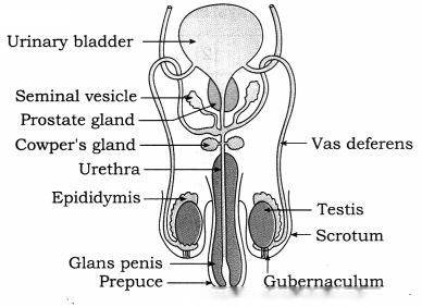
(ii) Testes:
(iii) Accessory ducts : Rete testis, vasa efferentia, epididymis, vas deferens, ejaculatory duct and urethra together form the accessory ducts of male reproductive system.
1. Vasa efferentia : Vasa efferentia are 12-20 fine tubules. They arise from rete testis and end into the epididymis. The sperms from the testis are carried by these ducts to the epididymis.
2. Epididymis : Epididymis are long and coiled tubes having three parts, viz. caput, corpus and cauda epididymis. They are located on the posterior border of each testis. The sperms undergo maturation in epididymis.
3. Vasa deferentia:
4. Ejaculatory duct : About 2 cm long pair of ducts formed by joining of vas deferens and a duct of seminal vesicle are the ejaculatory ducts. Both ejaculatory ducts open into urethra near the prostate gland. Seminal fluid containing spermatozoa are carried by ejaculatory duct to the urethra.
5. Urethra : The male urethra provides a common passage for the urine and semen hence is also called urinogenital duct.
(iv) Accessory glands : Associated with male reproductive system are : (a) Seminal vesicles (b) Prostate gland and (c) Cowper’s or Bulbourethral glands. Every accessory gland has secretion which helps in functions of reproductive system.
(v) External genitalia : External genitalia consists of penis and scrotum.
1. Penis:
2. Scrotum : The scrotum is a pouch of pigmented skin arising from lower abdominal wall. It protects testes within it. Scrotum acts as thermoregulator. Testis are suspended in scrotum by spermatic cord.
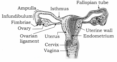
Internal organs are pair of ovaries and pair of fallopian ducts or oviducts, single median uterus and vagina. External genitalia is called vulva. There are a pair of vestibular glands in external genitalia. Mammary glands or breasts are also associated with reproductive system of female.
(1) Ovaries:
(2) Fallopian tubes/oviducts:
(i) Fallopian tubes lie horizontally over peritoneal cavity. These are about 10 to 12 cm long, narrow, muscular structure lined by ciliated epithelium.
(ii) They transport the ovum after ovulation from the ovary to the uterus.
(iii) Fallopian tube can be subdivided into the following three parts:
(3) Uterus/Womb:
(i) Uterus is a pear-shaped, highly muscular, thick walled, hollow organ measuring about 8 cm in length, 5 cm in width and 2 cm in thickness.
(ii) Uterus has the following three parts : Fundus, Body or corpus and Cervix.
(iii) The cervix communicates above with the body of the uterus by an aperture, the internal os and with vagina below by an opening the external os.
(iv) Uterus has three-layered wall. These layers are:
(v) Uterus receives the ovum from fallopian tube. It develops placenta during pregnancy for the nourishment of foetus. At the time of parturition, it expels the young one at birth.
(4) Vagina:
(5) External genitalia or vulva or pudendum : The external genitalia consists of five parts; viz. labia majora, labia minora, mons veneris, clitoris and vestibule.
(6) A pair of vestibular glands / Bartholin’s glands : These glands open into the vestibule and release a lubricating fluid.
(7) A pair of mammary glands/breasts : These are the accessory organs of female reproductive system for production and release of milk after parturition.
(2) In human beings fertilization is internal. Sperms deposited in vagina, swim across the uterus and fertilize the ovum in ampulla of the fallopian tube.
(3) Fertilization involves the following events:
(i) Insemination : Discharge of semen into the vagina at the time of copulation is called insemination.
(ii) Movement of sperm towards egg : Sperms reaching the vagina undergo capacitation process for 5-6 hours. During capacitation acrosomal membrane of sperm becomes thin and Ca++enters the sperm making it extra active. Sperms reach up to the ampulla by swimming aided with contraction of uterus and fallopian tubes. These contractions are stimulated by oxytocin of female. By capacitation sperm can reach ampulla within 5 minutes, they remain 5 viable for 24 to 48 hours, whereas ovum remains viable for 24 hours.
(iii) Entry of sperm into the egg : Though many sperms reach the ampulla, only a single sperm fertilizes the ovum. The acrosome of sperm after coming in contact with the ovum, releases lysins; hyaluronidase and corona penetrating enzymes. Due to these enzymes cells of corona radiata are separated and dissolved. The sperm head then passes through zona pellucida of egg. The zona pellucida has glycoprotein fertilizin receptor proteins. These bind to specific acid protein-antifertilizin of sperm. This makes sperm and ovum to come together. Fertilizin-Antifertilizin interaction is species- specific.
(iv) Acrosome reaction : When the sperm head comes in contact with the zona pellucida, its acrosome covering ruptures to release lytic enzymes, acrosin or zona lysin. These enzymes dissolve plasma membrane of egg so that the sperm nucleus and the centrioles enter the egg, while other parts remain outside. Now the vitelline membrane of egg changes into fertilization membrane which prevents any further entry of other sperms into the egg, thus polyspermy is prevented.
(v) Activation of ovum : After the entry of sperm head into ovum, it gets activated to resume and complete its meiosis-II. With this it gives out the second polar body. The germinal vesicle organises into female pronucleus. At this stage, it is true ovum.
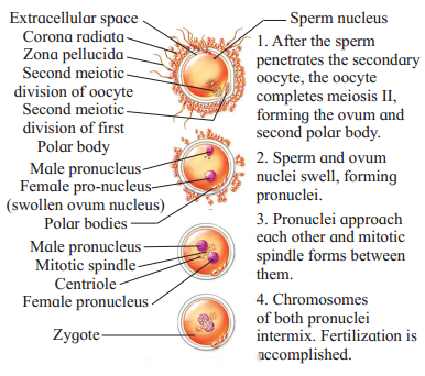
(vi) Fusion of egg and sperm : The coverings of male and female pronuclei degenerate results in the formation of a synkaryon by a process called syngamy or karyogamy. The zygote is thus formed.
(2) The type of cleavage in human is holoblastic, i.e. the whole zygote gets divided, radial and indeterminate, i.e. fate of each blastomere is not predetermined.
(3) Cleavage show faster synthesis of DNA and high consumption of oxygen.
(4) Since there is no growth phase between the cleavages, the size of blastomeres will be reduced with every successive cleavage.
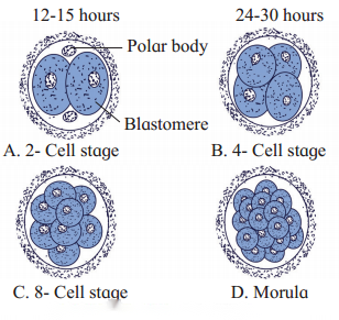
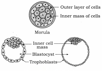
(5) The cleavages occur as follows:
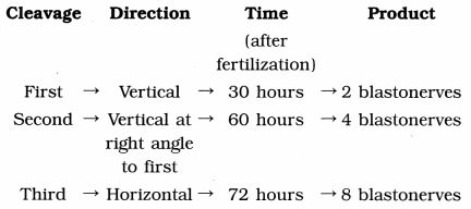
(6) Successive divisions produce a solid ball of cells called morula of 16 cells. It consists of an outer layer of smaller clearer cells and an inner mass of larger cells.
(7) Morula reaches the uterus about 4-6 days after fertilization.